
The Human Glymphatic System: Structure, FunctioImplicationsn, and Health 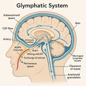
Overview of the Glymphatic System
The glymphatic system is a recently discovered waste-clearance pathway in the central nervous system (CNS) that parallels the lymphatic system in function. It consists of a network of perivascular channels, formed by glial cells (astrocytes), through which cerebrospinal fluid (CSF) circulates to flush out interstitial waste from the brain. In this system, CSF from the subarachnoid space enters the brain alongside arteries, moves through the brain tissue, and then exits alongside veins. The term “glymphatic” was coined in 2012 by neuroscientist Maiken Nedergaard to reflect the involvement of glia and its functional analogy to the lymphatic system. In essence, the glymphatic system serves as the brain’s own “plumbing system,” carrying fluid in and out of brain tissue in a highly organized way.
Figure 1: Diagram of the glymphatic system. Cerebrospinal fluid (CSF) flows from the subarachnoid space into the brain alongside arteries (left, red) via periarterial spaces (PVS) formed by astrocyte endfeet (yellow) containing water channels (AQP4). The CSF mixes with interstitial fluid (ISF) in the brain parenchyma, allowing exchange of solutes (clearance of waste like amyloid-β and tau, and distribution of nutrients). Fluid and waste then drain out along perivenous spaces (right, blue) toward arachnoid granulations and meningeal lymphatic vessels for elimination.
Components and function: A key feature of the glymphatic system is its use of glial cells to facilitate fluid transport. Astrocytes that surround brain blood vessels have specialized endfeet rich in aquaporin-4 (AQP4) water channels, which help drive CSF into the brain tissue. As arteries in the brain pulsate with each heartbeat, they “pump” CSF along these periarterial spaces and into the interstitium of the brain. There, the CSF mixes with interstitial fluid, picking up waste products. The fluid and waste then collect in perivenous spaces (around veins) and eventually drain out of the brain. Some of this fluid is funneled to conventional lymphatic structures: for example, it can exit via arachnoid granulations into the venous bloodstream or travel through recently discovered meningeal lymphatic vessels to lymph nodes. Importantly, the glymphatic circulation is not uniformly active at all times; it ramps up during sleep. During deep (non-REM) sleep, the brain’s interstitial space expands, resistance to fluid flow drops, and glymphatic clearance of toxins dramatically increases. In wakefulness, higher levels of neurotransmitters like norepinephrine cause the interstitial space to contract, reducing glymphatic flux – essentially putting the cleaning on “pause” while we are awake. This day-night difference suggests that one biological purpose of sleep is to allow the brain to enter a state that optimizes waste removal.
Role in Brain Health and Waste Clearance
The glymphatic system is crucial for maintaining the brain’s homeostasis by clearing metabolic waste and distributing essential compounds. It serves a function analogous to the lymphatic system in the rest of the body, which removes interstitial fluid and waste from tissues. In the brain, however, the glymphatic pathway handles this task. Studies have shown that glymphatic flow facilitates the removal of potentially neurotoxic proteins that accumulate during waking hours. For instance, the system efficiently clears amyloid-β and tau, the protein aggregates associated with Alzheimer’s disease, from the brain interstitium. It also aids in removing other metabolites and excess extracellular fluid, preventing harmful buildup. In addition to waste removal, the glymphatic network helps distribute nutrients and signaling molecules. Research indicates that compounds like glucose, lipids, and amino acids are disseminated through the brain via this convective CSF–interstitial fluid exchange mechanism. This dual role means the glymphatic system not only cleanses the brain but also nourishes it, supporting normal neural function.
Because the glymphatic system works most effectively during sleep, it underscores the link between sleep and brain health. During deep sleep, increased glymphatic activity flushes out waste products that accumulated during the day. Conversely, sleep deprivation or chronically poor sleep can impair this nightly cleansing. Human studies have found that even one night of sleep loss leads to less efficient clearance of amyloid-β and tau from the brain. Over time, such impairment may allow toxic substances to accumulate. This explains, in part, why long-term sleep problems are associated with cognitive decline and increased risk of neurodegenerative conditions. In short, a healthy glymphatic system—facilitated by regular deep sleep—is vital for overall brain health, preventing waste buildup that could interfere with neuronal function. It also appears to be influenced by cardiovascular health and lifestyle: factors like aging, hypertension, and exercise can affect glymphatic flow. Thus, the glymphatic system is emerging as a key piece in the puzzle of how systemic factors (sleep quality, heart health, etc.) tie into brain wellness.
Glymphatic Malfunction and Neurodegenerative Disease
When the glymphatic system fails to function properly, the brain’s ability to purge itself of waste is compromised. Such glymphatic dysfunction can lead to accumulation of neurotoxic debris, which is thought to set the stage for or exacerbate neurological diseases. A clear example is Alzheimer’s disease (AD). In AD, proteins like amyloid-β plaques and tau tangles build up in the brain; normally, the glymphatic system helps clear these species. Research indicates that a sluggish glymphatic system contributes to amyloid and tau accumulation. In animal models, disabling glymphatic pathways causes amyloid-β to linger much longer in brain tissue. Correspondingly, human studies using MRI proxies for glymphatic flow have found that AD patients have reduced glymphatic activity compared to healthy individuals. Notably, even in the preclinical or early stages of Alzheimer’s, lower glymphatic function (measured by an “ALPS index” on MRI) predicts faster buildup of amyloid plaques on PET scans. This suggests glymphatic impairment isn’t just a consequence of AD; it may be a contributing factor in its development. Consistent with this, common risk factors for dementia—aging, poor sleep, and vascular disease—are all known to impair glymphatic clearance. In fact, deep non-REM sleep (when glymphatic activity peaks) is often reduced in older adults and dementia patients, potentially accelerating the vicious cycle of waste accumulation.
A similar story is emerging in Parkinson’s disease (PD) and related disorders. PD is characterized by aggregates of α-synuclein protein (Lewy bodies) in the brain, and mounting evidence links this to glymphatic clearance issues. Patients with Parkinson’s have been observed to have diminished glymphatic flow on imaging studies, just like in AD. Lower glymphatic function in individuals with PD correlates with more rapid motor and cognitive decline, according to clinical assessments. This implies that an inefficient glymphatic system might allow toxic α-synuclein and other metabolites to accumulate, worsening neurodegeneration. Intriguingly, many PD patients experience sleep disturbances (especially loss of deep sleep and REM sleep behavior disorder) even before motor symptoms arise, and these sleep issues could further suppress glymphatic-mediated waste removal. The interplay of sleep, glymphatic function, and protein aggregation is an active area of research in PD.
Beyond AD and PD, glymphatic dysfunction has been implicated in a range of neurological conditions. Aging in general is associated with a decline in glymphatic efficiency – astrocytic AQP4 channels become less polarized to blood vessels in old animals, and the flow of CSF through the brain slows with age. This age-related decline may help explain why neurodegenerative diseases predominantly affect the elderly. Cerebral small vessel disease (CSVD), a condition of small blood vessel damage often seen in aging, correlates with impaired glymphatic function and cognitive impairment as well. In stroke, studies have found that glymphatic clearance on the stroke-affected side of the brain is reduced, possibly contributing to post-stroke edema and toxin buildup. Conversely, enhancing glymphatic activity is being explored as a way to improve stroke recovery. Traumatic brain injury (TBI) can acutely damage glymphatic pathways, and greater axonal damage in TBI has been associated with worse glymphatic dysfunction. Chronic traumatic encephalopathy (the neurodegenerative condition linked to repetitive head injuries) might also involve long-term glymphatic impairment. Normal pressure hydrocephalus (NPH), a disorder of CSF circulation, shows delayed clearance of fluid and waste on glymphatic imaging, suggesting a link between poor glymphatic drainage and the cognitive symptoms in NPH. Even multiple sclerosis and other inflammatory or demyelinating diseases are being examined for glymphatic involvement, given that immune cell traffic and waste clearance in the CNS could influence disease progression. The breadth of conditions connected to the glymphatic system is striking – from dementia, to stroke, to TBI, to sleep apnea – and underscores how fundamental this clearance mechanism is to brain health. Indeed, some scientists have gone so far as to propose that a failure of the glymphatic system may be a “final common pathway” leading to dementia and neurodegenerative disease. While multiple factors contribute to these diseases, a malfunctioning glymphatic system appears to exacerbate the pathology by allowing toxic waste to accumulate when it should be removed.
Recent Research and Discoveries (2024–2025)
Since its initial discovery just over a decade ago, research into the glymphatic system has been rapidly evolving. In 2024, a major breakthrough provided the most direct evidence yet of the glymphatic system in humans. For the first time, scientists were able to visualize CSF flow through the human brain’s perivascular spaces in living patients. In this study (published in PNAS in October 2024), five neurosurgery patients had a contrast agent injected into their CSF during tumor removal surgery. MRI scans taken hours later revealed the contrast-labeled CSF moving along specific pathways in the brain – tracing the outlines of arteries and dispersing through the brain tissue, then accumulating around veins. Rather than diffusing randomly like a sponge soaking up water, the fluid followed distinct channels consistent with the glymphatic network. “Nobody has shown it before now,” remarked the lead author, noting that this definitive visualization in humans answers long-standing skepticism about the glymphatic system’s existence. The study concluded that perivascular spaces do act as conduits for CSF flow in humans, directly supporting the glymphatic model. This discovery not only cements our understanding of human brain physiology, but also highlights new clinical opportunities. Knowing that the glymphatic pathway is active in people suggests it could be targeted for improving brain waste clearance. The authors of the 2024 study emphasized that their findings support pursuing lifestyle measures and therapies to “maintain and enhance” glymphatic function in humans. In other words, keeping this brain plumbing flowing—through good sleep habits, cardiovascular health, or even new medications—might help stave off neurological diseases. This has spurred interest in practical ways to monitor and boost glymphatic activity in patients.
Researchers are now developing noninvasive imaging techniques to assess glymphatic function routinely. One approach uses specialized MRI sequences and diffusion tensor imaging to measure the movement of water along white matter tracts adjacent to perivascular spaces. The resulting metric, known as the “ALPS index,” reflects glymphatic efficiency in certain brain regions. In the last couple of years, many studies have applied the ALPS index (and other MRI methods) in various populations – and their results reinforce the clinical importance of glymphatic flow. A recent meta-analysis (2023) combining results from 11 studies in AD and 12 studies in PD found strong evidence of reduced glymphatic function in both Alzheimer’s and Parkinson’s patients compared to controls. Lower ALPS values in these individuals were linked to greater disease severity, such as more amyloid in the brain or faster motor decline. These findings validate the glymphatic system’s role in human disease and even hint at its potential as a biomarker: one could imagine using an MRI-based glymphatic index to identify people at risk for dementia before symptoms arise, or to monitor the efficacy of interventions aimed at improving waste clearance.
Other new research is exploring how to modulate the glymphatic system for therapeutic benefit. Because glymphatic clearance is tied so closely to sleep, scientists are examining whether enhancing deep sleep can in turn enhance waste removal. For example, trials have looked at acoustic stimulation – playing certain sounds to amplify slow-wave (delta) brain activity during sleep – to see if it boosts glymphatic exchange. There are also investigations into pharmacological avenues: since astrocytic AQP4 water channels are critical for glymphatic flow, drugs that upregulate AQP4 or improve astrocyte function might accelerate clearance. Conversely, some researchers are asking if blocking glymphatic pathways could ever be beneficial (for instance, to prevent the spread of toxic proteins after acute injury), though the focus is largely on enhancement. Another intriguing line of inquiry is the effect of body posture and daily habits on glymphatic circulation. Studies in animals and humans found that sleeping in the lateral position (on one’s side) clears brain waste more effectively than sleeping on the back or stomach. This has led to recommendations for side-sleeping as a simple way to potentially improve nightly glymphatic function. Meanwhile, neuroscientists are delving into the cellular-level regulation of glymphatic flow: a 2023 study showed that light stimulation of specific brain circuits in mice could improve glymphatic clearance and reduce Alzheimer-like pathology, pointing to possible neuromodulation strategies. In sum, as of 2024–2025, the glymphatic field is moving fast – transitioning from basic discovery to translational research. Ongoing studies are defining how factors like exercise, diet, and brain stimulation impact this clearance system. The hope is that by understanding and harnessing the glymphatic system, we can develop novel ways to prevent neurodegeneration, improve recovery from brain injuries, and overall keep the “housekeeping” of the brain running optimally.
Glymphatic System vs. Lymphatic System: Key Comparisons
The glymphatic system is often described as the brain’s equivalent of the body’s lymphatic system. Both systems handle fluid balance and waste removal, but they operate in different contexts and with distinct anatomy. Below are the key differences and connections between the glymphatic and lymphatic systems:
- Location and Structure: The lymphatic system is a body-wide network of vessels and nodes that collects excess interstitial fluid from tissues and returns it to the bloodstream. These lymphatic vessels are lined with endothelial cells and have one-way valves, and lymph nodes serve as filtration and immune centers along the path. In contrast, the glymphatic system is confined to the brain (and spinal cord) and does not have separate vessels in the parenchyma. Instead, it piggybacks on the brain’s vasculature: CSF flows through periarterial spaces and perivenous spaces created by astrocyte endfeet, effectively using the spaces around blood vessels as channels. There are no lymph nodes in brain tissue; waste is cleared directly through fluid flow into drainage pathways.
- Fluid and Driving Force: The fluid in the lymphatic system, called lymph, originates as interstitial fluid that enters lymphatic capillaries. Its movement is driven by muscle contractions, body movements, and intrinsic vessel contractions; valves in lymph vessels ensure one-way flow toward the heart. The glymphatic system, on the other hand, moves cerebrospinal fluid which mixes with interstitial fluid in the brain. Glymphatic flow is driven predominantly by arterial pulsation (the rhythmic beating of arteries pushes the fluid) and is facilitated by the pumping action of astrocytes (via AQP4 water channels). Unlike lymph, which flows continuously albeit slowly, glymphatic circulation is heavily dependent on the sleep-wake cycle – it pulses during sleep and is comparatively reduced during wakefulness. In summary, lymphatic flow relies on mechanical forces and vessel muscles, whereas glymphatic flow relies on cardiovascular pulsatility and neural state (sleep) to propel fluid.
- Immune Function: The lymphatic system is a critical component of the immune system. Lymphatic vessels transport white blood cells (like lymphocytes) throughout the body and carry antigen-presenting cells to lymph nodes, where immune responses are triggered. This immune surveillance function means the lymphatic system not only removes waste but also scans it for pathogens. The glymphatic system is not known for actively transporting immune cells within the brain tissue (the brain has its own resident immune cells, microglia, for local surveillance). However, it does have an immune connection: by draining CSF and interstitial fluid to the peripheral lymphatic system, the glymphatic pathway can deliver brain-derived antigens and any immune cells or cytokines in the CSF to the lymph nodes. In fact, in 2015 researchers discovered meningeal lymphatic vessels in the dura mater (outer lining of the brain) that directly connect the glymphatic outflow to the body’s lymph nodes. These meningeal lymphatics carry CSF and immune cells from the subarachnoid space and drain into the deep cervical lymph nodes, thereby linking brain waste clearance with the immune system. This was a groundbreaking finding, as it showed the brain is not completely isolated from the lymphatic network – there is a point of interface where glymphatic-cleared fluid is handed off to conventional lymphatics.
- Unique Role and Integration: In the rest of the body, the lymphatic system handles not only waste removal but also fat absorption from the gut and returning plasma proteins to circulation. The glymphatic system is specialized solely for the CNS environment, where tight skull confines and the blood–brain barrier necessitated a different solution for waste clearance. One can think of glymphatic pathways as a functional extension of the lymphatic system into the brain’s territory, using a clever design of existing structures (perivascular spaces and glial cells) to perform lymphatic-like duties. Ultimately, the glymphatic and lymphatic systems converge: fluid and solutes cleared by glymphatic flow exit the brain and enter lymphatic vessels or the bloodstream, becoming part of the overall circulatory and excretory system of the body. In summary, the lymphatic system is a traditional vessel-based drainage network throughout the body, whereas the glymphatic system is a glia-supported perivascular clearance mechanism within the brain. Both are interdependent – the glymphatic system feeds into lymphatic routes – and together they maintain the delicate fluid and waste balance necessary for our tissues, especially the highly sensitive brain, to function properly.
-
The Perrin Technique and the Glymphatic System
The Perrin Technique, developed by Dr Raymond Perrin, has been used for years to stimulate lymphatic and glymphatic drainage. It combines manual therapy, spinal alignment, and lymphatic massage to help the body clear brain and spinal toxins. While it originally gained attention for treating chronic fatigue and ME/CFS, it is now gaining traction for its alignment with emerging glymphatic research. We have been practising the Perrin Technique here at the Willows Clinic for nearly two decades.
Conclusion: The discovery of the glymphatic system has fundamentally changed our understanding of brain physiology. It revealed that the brain, once thought to lack a direct waste removal system, actually possesses a specialized “cleaning service” akin to lymphatics. This system’s importance is now evident: it keeps brain cells healthy by clearing toxins, and its dysfunction is linked to diseases like Alzheimer’s and Parkinson’s. Cutting-edge research up to 2025 has confirmed the glymphatic pathway in humans and opened new avenues to diagnose and possibly treat neurological diseases by targeting brain waste clearance. As we continue to unravel the glymphatic system’s workings and its interplay with the lymphatic and other systemic systems, we move closer to therapies that could enhance this nightly brain cleansing. Ensuring the glymphatic system runs smoothly – through healthy sleep, lifestyle, or future medical interventions – may prove to be a key strategy in preserving cognitive function and preventing neurodegeneration.